Color the arrow indicating mitosis purple. Reset Late Proplase Cytokinesis Telophase 253 PM di.
Mitosis And Cytokinesis Teaching Resources Teachers Pay Teachers
View Art-labeling Activity - Interphase Mitosis and Cytokinesisjpg from BSC MISC at Miami Dade College Miami.

. Students describe what happens at each stage. Reproduction production of gametes Looking through a light microscope at a cell undergoing meiosis you see that the chromosomes have joined into XX-shaped tetrads. Worksheet for activity 2 from nih.
1 sample diffusion 2 facilitated diffusion 3 filtration 4 osmosis 5 active transport 6 endocytosis 7 exocytosis A. Mitosis and Meiosis Online Lab Name. Which statement best describes strand characteristics as it relates to DNA replication.
View Lab Report - Mitosis and Meiosis Online Lab from BSC 1041 at Miami Dade College Miami. This activity includes 10 questions covering the process and phases of mitosis and the stages of the cell cycle - interphase prophase metaphase anaphase telophase and cytokinesis. The cell membrane engulfs a particle or substance drawing it into the cell in a vesicle B.
Harold has psoriasis a skin disorder that speeds up the life cycle of the skin cells. These tetrads are lined up along a plane that runs through the center of the cell. The function s of meiosis isare _____.
Art-labeling Activity - Interphase Mitosis and Cytokinesisjpg School Miami Dade College Miami Course Title BSC MISC Uploaded By Squirtle91 Pages 1 Ratings 100 2 2 out of 2 people found this document helpful This preview shows page 1 out of 1 page. Walking dead photo gallery. The blastocyst has 2 distinct cell populations.
Word bank for the above. Interphase G2 Mitosis Mitotic Phase. What moves the chromatids during mitosis.
The chromosomes line up separately on the spindle for metaphase of mitosis but at the first division of meiosis the chromosomes pair in a process called synapsis. Chamberlain liftmaster elite series battery replacement. During which phase is the dna duplicated in mitosis.
Figure 331 Drag the appropriate labels to their respective targets. View art labeling activity - ventricles of the brain Lateral viewjpg from BSC MISC at Miami Dade College Miami. Use the recitation PPT slides alongside this worksheet.
Feb 12 2015 - This 16 piece science clip art set features 8 phases of cell mitosis from interphase to cytokinesis. Comparison of Meiosis and Mitosis There are two divisions instead of one. Students who viewed this also studied Mitosis and Meiosis Online Lab lab 4.
No canvas element supported The cell cycle can be described in several ways. View Notes - Lecture 34 - Mitosis and Cytokinesispdf from BIOL 200 at University of British Columbia. Marengo il police reports.
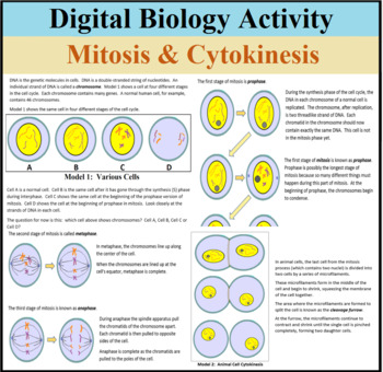
Which of the following builds new strands of DNA. 1 of 2 Art-labeling Activity. Mitosis Worksheet - Label each stage and Exam-style Qs by learnwithlaura 120 Zip A PDF worksheet focusing on the 4 stages of mitosis.
Theyll use one website to read about the general cell cycle with a focus on the cells. Match the movements into and out of the cell on the left with their descriptions on the right. S G1 Cytokinesis Right.
Phases of the Cardiac Cycle Label the sequence of events that occur during a cardiac cycle. Nucleus nuclear envelope nuclear pore plasma membrane cytosol rough. Unit 7 Lecture 4 December 1 2017 Unless otherwise indicated ALL BIOL200 Material.
Label the following diagram of mitosis of an animal cell. Diagram that shows the phases of the cell cycle. Meiosis labeling activity answer key.
Can be used as a formative assessment or independent -learning taskAnswer sheet provided. Then drag the blue labels onto the blue targets to identify the key stages that occur during those phases. This cell is in _____.
Part A - The cell cycle Drag the pink labels onto the pink targets to identify the two main phases of the cell cycle. A g1 phase b. Mitosis worksheet diagram identification.
Movement down a concentration gradient through an ion channel or with. Label each of the following drawings of cells in different stages of mitosis and cytokinesis. There are exam-style questions to check student understanding.
Figure 32 2 of 2 Which of the following is NOT one of the three major components of a typical eukaryotic cell. The cell prepares for mitosis. To review a crucial phase of the cell cycle watch this BioFlix animation.
There is also a 4 page graphic organizer chart that includes a drawing of each of the organelles in alphabetical order. Start studying Art-labeling Activity. Oishii ramen and poke ingleside.
Nucleus Ribosome Ribosomes are organelles and they are located within one of the major components of the cell. The cytoplasm divides the cell membrane pinches inward ultimately producing two daughter cells.
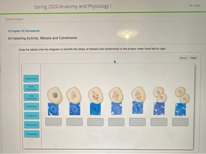
Solved Spring 2020 Anatomy And Physiology Hi Lubia Course Chegg Com
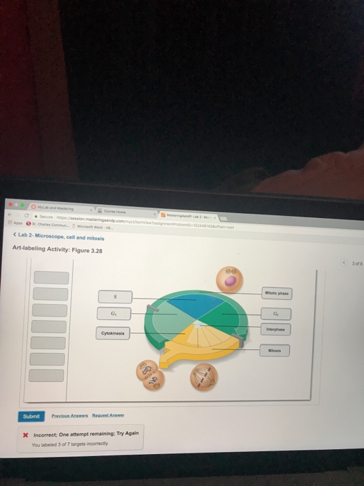
Solved Corse さ Ore C Lab 2 Microscope Cell And Mitosis Chegg Com
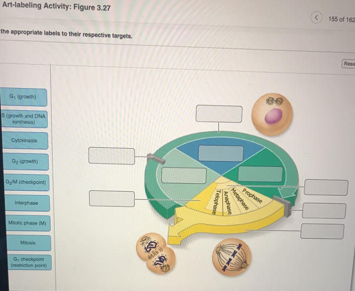
Solved Art Labeling Activity Figure 3 27 155 Of 162 The Chegg Com
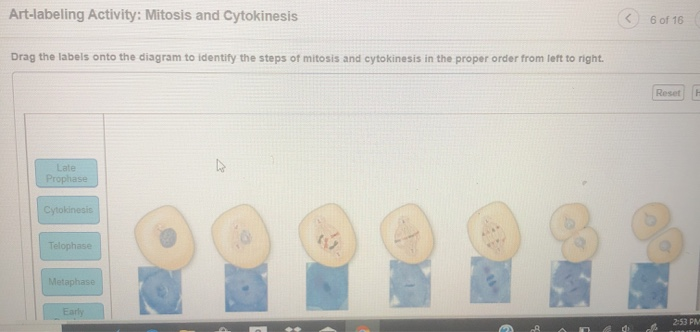
Solved Art Labeling Activity Mitosis And Cytokinesis 6 Of Chegg Com

Cell Division Mitosis Artwork And Labels Free Teaching Resources
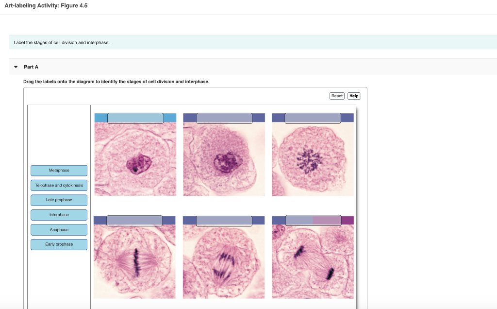
Solved Art Labeling Activity Figure 4 5 Label The Stages Of Chegg Com
Mitosis And Cytokinesis Cell Labeling Key By K I S S It Biology Tpt

Chapter 1 2 Lab Flashcards Quizlet
0 comments
Post a Comment